
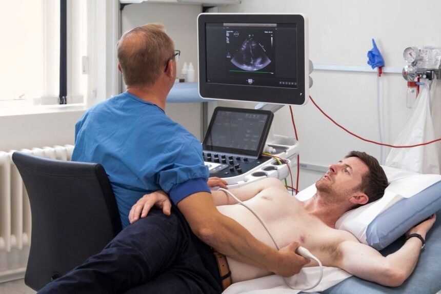
Body imaging refers to a group of noninvasive techniques that create detailed pictures of the structures inside your body. Using tools such as computed tomography (CT), magnetic resonance imaging (MRI), and ultrasound, radiologists can examine organs, blood vessels, and tissues without surgery. These images allow specialists to examine areas that a standard physical exam cannot reach, providing a clearer view of internal health. A radiologist may recommend body imaging when there is a need to observe the heart and blood vessels in greater detail. Here’s information on body imaging and how it can help detect cardiovascular disease early:
How Does Body Imaging Work?
Family history, existing risk factors, or symptoms noted by a physician can all lead to a body imaging referral. Radiologists interpret the resulting scans, and their findings help your care team understand your cardiovascular system. Different imaging methods rely on different forms of energy to produce their pictures. CT scans use X-rays taken from multiple angles, which a computer combines into cross-sectional images. MRI uses strong magnetic fields and radio waves to map soft tissues, while ultrasound sends sound waves into the body and records the echoes that bounce back.
Each method offers unique strengths. CT captures fine detail of bones and blood vessels quickly, and MRI provides excellent contrast between soft tissues. Ultrasound is portable and does not use radiation, so it is often selected for repeated examinations. The choice of technique depends on the area being studied and the information a radiologist needs to gather.
What Are Imaging Cardiovascular Assessments?
Imaging cardiovascular assessments are scans focused specifically on the heart and the network of blood vessels. These examinations measure structure, blood flow, and the presence of substances such as calcium within arterial walls. The results give physicians objective data to review alongside other test findings.
Coronary Calcium Scoring
A coronary calcium scan uses CT to detect calcium deposits in the heart’s arteries, providing live images of heart function. The scan produces a numerical score based on the amount of calcium present. Radiologists report this score, and physicians use it as one factor when assessing a patient’s cardiovascular profile.
Cardiac MRI and CT Angiography
Cardiac MRI examines the size, shape, and movement of the heart’s chambers and walls. CT angiography, by contrast, focuses on the blood vessels and can show narrowing or blockages. Both approaches deliver detailed images that support a thorough evaluation of the cardiovascular system.
When May You Need One?
A physician may recommend cardiovascular imaging if you have risk factors that warrant closer monitoring. High blood pressure, elevated cholesterol, diabetes, or a family history of heart conditions are common reasons for a referral. In these situations, imaging offers a way to gather additional detail before symptoms progress. Age and lifestyle can also influence the decision to assess your cardiovascular system. Your radiologist can weigh your personal history against clinical guidelines and decide whether imaging or another assessment type is appropriate.
Get Cardiovascular Body Imaging
Cardiovascular body imaging gives your care team a detailed view of your heart and blood vessels, supporting informed conversations about your health. If you have cardiovascular risk factors, speak with your provider about which body imaging option is appropriate for your situation. A radiologist can explain what to expect during different examinations and how the images will be obtained and reviewed. Scheduling an assessment often begins with a referral from your primary physician, who will coordinate the details with an imaging center.
- Exploring the Long-term Effects of Varicose Veins
- Advances in Body Imaging for Early Detection of Cardiovascular Disease
- Understanding the Limitations of Disc Replacement Surgery
- How to Prepare for a Skin Cancer Screening With Your Dermatologist
- What You Need To Know About Laser Eye Surgery for Astigmatism